
Page 2 /
Good quality with appropriate penetration, little rotation and adequate inflation. Soft tissues and bony structures normal. Normal cardiac silhouette. Normal appearance of trachea and mainstem bronchi. Lung fields clear.
The patient was born at term following a normal pregnancy, and had a normal perinatal course. There was no personal history of allergies or eczema. He did have a history of chronic nasal obstruction for 6-24 months according to various reports. He had been treated for episodes of sinusitis in the past year, and had had tympanostomy tubes placed 2 months prior to presentation for chronic otitis media with effusion complicated by conductive hearing loss. He was in Grade 7 with good school performance, and had been quite active, with participation in many sports. Family history was positive for otosclerosis, but was otherwise unremarkable.
On examination in the emergency department:
Respiratory rate 24, Heart rate 100, Blood pressure 101/66, SaO2 95% on R/A, T 37.1oCs
Weight 60.3kg (75-90%ile), Height 171cm (75-90%ile)
Looks well; alert; cooperative; no distress; muffled, hoarse voice
Head and neck No cervical/axillary lymphadenopathy; tympanic membranes's - tympanic-tubes bilaterally; oropharynx normal
Chest: pectus excavatum, no increased work of breathing
Good air entry bilaterally
Stridor at rest, worse on forced inspiration; no other adventitious sounds
Cardio-vascular system: normal
Abdomen: normal
A soft tissue neck x-ray was ordered in the Emergency department.
 Next page /
ANSWER
Good quality with appropriate penetration, little rotation and adequate inflation. Soft tissues and bony structures normal. Normal cardiac silhouette. Normal appearance of trachea and mainstem bronchi. Lung fields clear.
The patient was born at term following a normal pregnancy, and had a normal perinatal course. There was no personal history of allergies or eczema. He did have a history of chronic nasal obstruction for 6-24 months according to various reports. He had been treated for episodes of sinusitis in the past year, and had had tympanostomy tubes placed 2 months prior to presentation for chronic otitis media with effusion complicated by conductive hearing loss. He was in Grade 7 with good school performance, and had been quite active, with participation in many sports. Family history was positive for otosclerosis, but was otherwise unremarkable.
On examination in the emergency department:
Respiratory rate 24, Heart rate 100, Blood pressure 101/66, SaO2 95% on R/A, T 37.1oCs
Weight 60.3kg (75-90%ile), Height 171cm (75-90%ile)
Looks well; alert; cooperative; no distress; muffled, hoarse voice
Head and neck No cervical/axillary lymphadenopathy; tympanic membranes's - tympanic-tubes bilaterally; oropharynx normal
Chest: pectus excavatum, no increased work of breathing
Good air entry bilaterally
Stridor at rest, worse on forced inspiration; no other adventitious sounds
Cardio-vascular system: normal
Abdomen: normal
A soft tissue neck x-ray was ordered in the Emergency department.
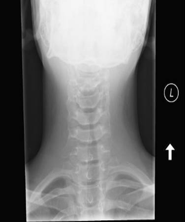 Figure 2a |
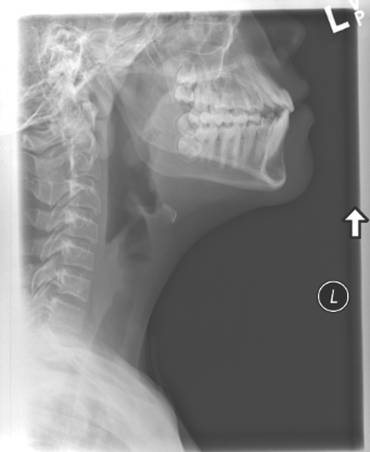 Figure 2b |
PLEASE DESCRIBE THE X-RAY?